2. ER에서 해야 할 처치
- ABC 유지
Airway 유지 - intubation
Breathing and ventilation
Circulation
- IICP 방지
head elevation
sedation
osmotic agents
- Brain CT
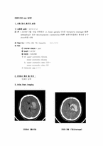
3. Initial brain imaging
- brain CT : ventricle에 moderate amount의 high density hemorrhage가 관찰되며
midbrain과 right basal ganglion의 lenticular nucleus에 h
2. ER에서 해야 할 처치
1) ABCDE 유지
Airway 유지 - intubation
Breathing and ventilation
Circulation
disability
exposure
2) Brain CT, C-spine lateral
3. Initial Brain Imaging
1) C- spine lat. : fracture in C5 vertebral body with complete dislocation.
2) C- spine CT, 3D
4. 향후 Plan & 보호자에게 말해 줄 수 있는 Complicat
(1) X-ray
진단 어려움.
정상적인 척추전만(lordosis)의 소실, 추간판 공간의 감소가 관찰가능하나 비특이적.
추간판공간의 감소가 있는 경우에는 추간판에 연한 척추체의 endplate가 정상이거나 약간의 퇴행성 변화를 보임.
(2) MRI –명랑군 case 분석에의 적용
1) Mid-sagittal image
CSF는 수분이 많고 disc는
name : Ventriculo-peritoneal shunt
3) OP 한 이유 : Enlarged Rt. lateral ventricle
6. F/U CT
1월 19일 1월 26일
7. 2nd OP
1) OP date : 2010/2/1
2) OP name : cranioplasty with bone cement
3) OP 한 이유 : skull defect(Lt.)
8. F/U CT
2월 2일 3월 2일
의한 증상: Anterior Cerebral artery와 Corpus callosum 손상
수술에 의한 합병증: 감염- 수막염 및 뇌 농양
5. 1st OP
OP date : 2010/02/27
OP name : craniectomy,Lt.
OP 한 이유 : 감압 및 swelling되는 brain tissue 보호하기 위해
6. F/U CT
Tx: Craniectomy&subdural catheter(왼쪽 CT에서 brain contusion 보임)
4. 향후 Plan & 보호자에게 말해 줄 수 있는 Complication
- IV urokinase for thrombolysis
- Complication
뇌출혈
기타 장기의 출혈
출혈로 인한 사망
조영제에 의한 s/e
색전증에 의한 다른 문제
5. IA uk 후 problem
- 환자 시술 당일 저녁 mental change 보여서 f/u CT 촬영
6. F/U brain imaging
<1/19 f/u CT>
3. Initial Brain Imaging
1) angio
2) CT
4. 향후 Plan & 보호자에게 말해 줄 수 있는 Complication
- 뇌혈관조영술, 절대안정, 코일 색전술, 결찰
- complication : 재출혈, 뇌연축, 수두증
- 수술에 대한 complication : vomitting, 경련, shock
5. 1st OP
1) OP date : 2010/02/24 수술예정
2) OP name :
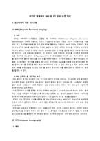
History, Clinical features, CT, Angiography, MRA, MRV, Biopsy etc.
Emergent noncontrast cranial CT
Standard initial imaging technique
DDx from ICH or mass lesion
Identify parenchymal bleeding >1cm in diameter
Hyperdense artery sign (acute thrombus in a vessel)
Sulcal effacement
Loss of the insular ribbon
Loss of gray-white interface, mass effect
Acute hypodensity
Image study
Chest PA(04.18) :
No newly developed lesion to suggest metastasis or active lung parenchymal disease.
Abdomen Flat, Upright(04.18) : Normal
CT Abd+Pel Dynamic (04.18)
Rectal cancer with liver metastases post-op, s/p RFA.
Loculated fluid collection in the anterior aspect of Lt. lobe of the liver.
No evidence o